
Magnetic resonance diffusion tensor imaging (DTI), a novel imaging and post-processing technique developed on the basis of diffusion-weighted imaging (DWI), is currently the only method can reveal non-invasively unique information of white matter fiber tracts in the brain.
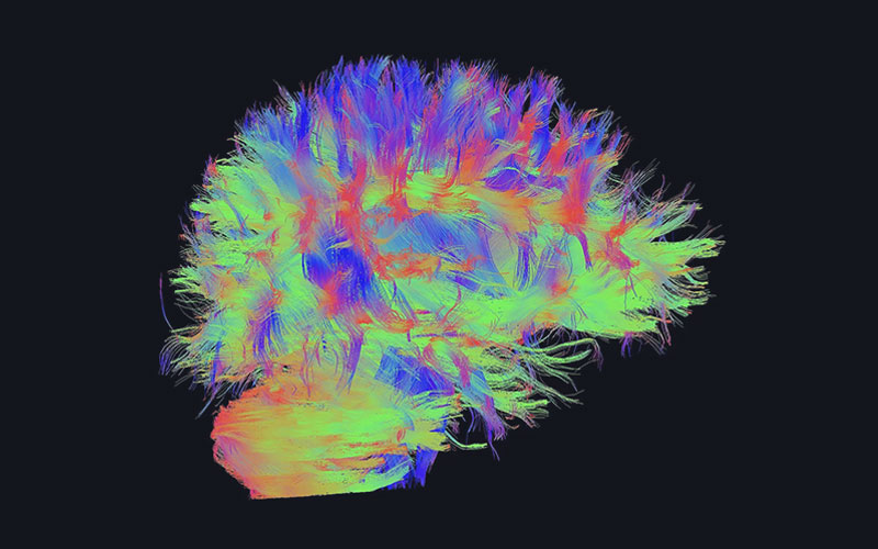
Simply put, the diffusive movement of water molecules in different tissues is affected by various tissue characteristics such as tissue type, integrity, structure and tissue barrier. The basic concept of DTI is that by observing the diffusive movement of water molecules in different directions in the same tissue, the anisotropy of water molecule diffusion in that tissue (i.e., different rates in different directions) can be obtained, thus reflecting the relevant characteristics of the measured tissue. For example, the diffusion of water molecules in the white matter (WM) of the brain presents less obstructions along the axon direction, but there is a greater restriction of diffusion in the radial direction, so the rate of diffusion of water molecules along the axon direction is higher. Based on this feature, DTI can measure the direction-dependent properties of water molecule diffusion in the white matter of the brain, and thus characterize the direction of the white matter fibers and trace the white matter fiber bundles throughout the brain. The diffusion behavior of water molecules in cerebral gray matter (GM) and cerebrospinal fluid (CSF) trends to be isotropic and shows a low signal on the DTI anisotropy score map.

The imaging quality of DTI is easily affected by various factors such as the homogeneity of the main magnetic field, gradient field performance, eddy currents, etc. Therefore, it has extremely high requirements on the MR hardware system, imaging sequence and related software system. However, at Wandong, all the challenges in DTI imaging have been overcome one by one.
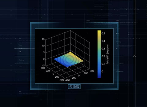
No matter it is 71cm large aperture system or 60cm aperture system, we control the magnetic field homogeneity in the core field for fat-suppression to the industry's top level. We also reduce geometric distortion and magnetization artifacts caused by Bo field inhomogeneity through ultra-fast pre-scan shimming technology.
[Unfold]
No matter it is 71cm large aperture system or 60cm aperture system, we control the magnetic field homogeneity in the core field for fat-suppression to the industry's top level. We also reduce geometric distortion and magnetization artifacts caused by Bo field inhomogeneity through ultra-fast pre-scan shimming technology.
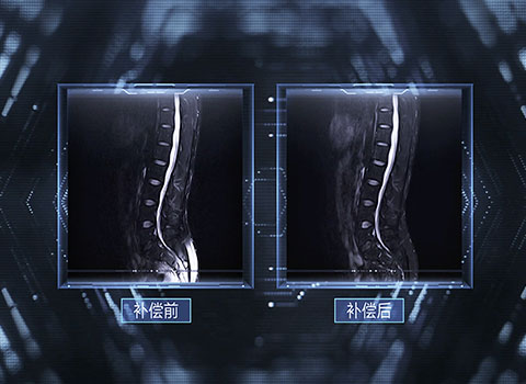
DTI generally uses the echo planar imaging (EPI) method for data acquisition. EPI acquisition is very sensitive to eddy currents, especially short eddy currents that can cause Nyquist artifacts. Wandong's eddy current compensation technology can minimize eddy currents in all spatial and temporal scales, minimizing DTI image artifacts and distortions.
[Unfold]
DTI generally uses the echo planar imaging (EPI) method for data acquisition. EPI acquisition is very sensitive to eddy currents, especially short eddy currents that can cause Nyquist artifacts. Wandong's eddy current compensation technology can minimize eddy currents in all spatial and temporal scales, minimizing DTI image artifacts and distortions.

The use of all-digital fiber-optic RF technology ensures that the DTI with a high enough signal-noise-ratio to achieve high-resolution imaging, allowing for excellent isotropic voxel imaging on 1.5T superconducting systems (with in-plane resolution and layer thickness of the same size, e.g., 2 × 2 × 2.5 mm3).
[Unfold]
The use of all-digital fiber-optic RF technology ensures that the DTI with a high enough signal-noise-ratio to achieve high-resolution imaging, allowing for excellent isotropic voxel imaging on 1.5T superconducting systems (with in-plane resolution and layer thickness of the same size, e.g., 2 × 2 × 2.5 mm3).

Provides high gradient field strength of 35mT/m and a gradient slew rate comparable to 3T magnet at 175mT/m/ms, supporting EPI with shorter echo intervals, thereby reducing sensitivity to motion, and reducing the effects of geometric distortion and image blurring.
[Unfold]
Provides high gradient field strength of 35mT/m and a gradient slew rate comparable to 3T magnet at 175mT/m/ms, supporting EPI with shorter echo intervals, thereby reducing sensitivity to motion, and reducing the effects of geometric distortion and image blurring.
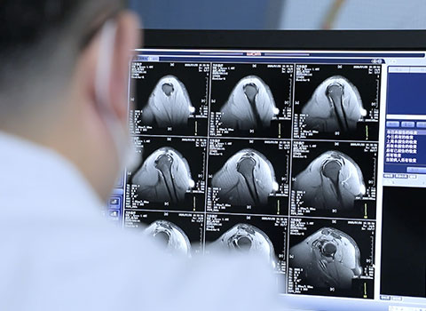
A new parallel imaging acquisition mode combining phase encoding inversion and field map correction with a high channel RF coil can eliminate the geometric distortion caused by magnetization artifacts and B0 field inhomogeneity, resulting in fantastic DTI images. In terms of data processing, artifacts and noise are further removed from the images by quality control and pre-processing, providing consistency for reliable tensor estimation.
[Unfold]
A new parallel imaging acquisition mode combining phase encoding inversion and field map correction with a high channel RF coil can eliminate the geometric distortion caused by magnetization artifacts and B0 field inhomogeneity, resulting in fantastic DTI images. In terms of data processing, artifacts and noise are further removed from the images by quality control and pre-processing, providing consistency for reliable tensor estimation.
Wandong's DTI data analysis function can provide all DTI parameter metrics (Figure 1), such as mean diffusivity (MD) or apparent diffusion coefficient (ADC), exponentiated apparent diffusion coefficient (eADC), axial diffusivity (AD), radial diffusivity (RD), volume ratio (VR), fractional anisotropy index (FA), and color FA map.
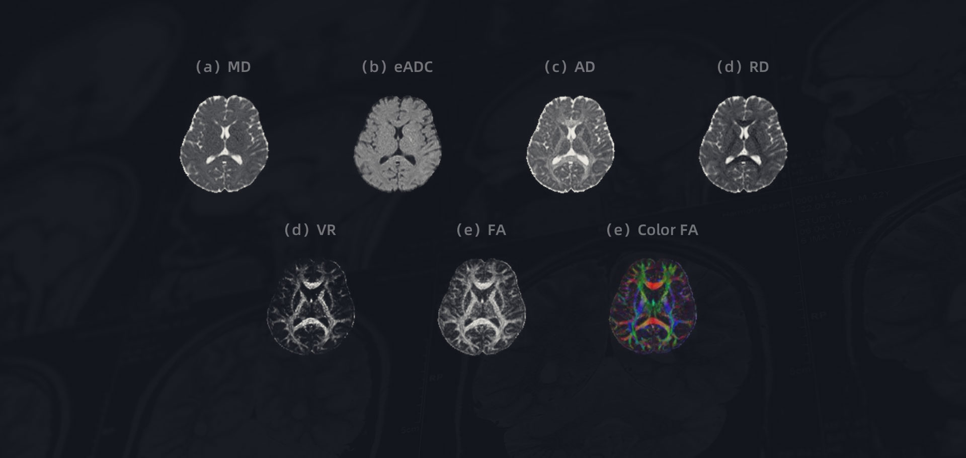
Parameter metrics after DTI data analysis and processing:
(a)mean diffusivity MD (b)exponentiated apparent diffusion coefficient eADC (c)axial diffusivity AD (d) fractional anisotropy index FA (e)color FA map
One of the biggest challenges of DTI is to present tensor information in an intuitive and easy-to-understand manner, most typically by forming 3D fiber bundle tracking maps, similar to those in color FA maps, with red representing left-to-right diffusion direction, green representing back-to-front, and blue representing bottom-to-top diffusion. Wandong's DTI data analysis function assigns fibers by continuously tracking specific anatomical tracts defined based on ROIs, thus enabling a comprehensive exploratory visualization of tensor data across the brain.

Nerve fiber bundles in coronal, sagittal and axial positions after DTI data analysis and processing
Special researcher at the Biomedical Imaging Research Center, Department of Biomedical Engineering, Tsinghua University School of Medicine, with research interests including high spatial and temporal resolution magnetic resonance neuroimaging technology development, quantitative imaging of in biopsy pathology information based on magnetic resonance images, etc.
And that is just the beginning.

1 Soares JM, Marques P, Alves V and Sousa N (2013) A hitchhiker's guide to diffusion tensor imaging. Front. Neurosci. 7:31. doi: 10.3389/fnins.2013.00031
2 Yiping Lu, Xuanxuan Li, Daoying Geng, et al. Cerebral Micro-Structural Changes in COVID-19 Patients -- An MRI-based 3-month Follow-up Study. ECLINICALMEDICINE 25: 100484 (2020).
3 Xiong Y, Li G, Dai E, Wang Y, Zhang Z, Guo H. Distortion correction for high-resolution single-shot EPI DTI using a modified field-mapping method. NMR Biomed. 2019 Sep;32(9):e4124. doi: 10.1002/nbm.4124. Epub 2019 Jul 4. PMID: 31271491.
 WeChat
 Twitter
 Linkedin
Contact Us
010-84575792/3/5/6
international@wandong.com.cn